Getting Started with Spatial Transcriptomics: Key Considerations Before You Begin
Spatial transcriptomics represents a cutting-edge methodology for profiling gene expression within preserved tissue sections, maintaining the native spatial context of cells and their surrounding microenvironments. In contrast to bulk RNA sequencing and even single-cell RNA-seq, spatial transcriptomics generates spatially resolved transcriptome maps, enabling high-resolution visualization of transcriptional heterogeneity across tissue architecture.
By delineating cell-type composition, spatial gene expression gradients, and intercellular signaling networks, this approach provides nuanced insights into tissue physiology and pathobiology. It has emerged as a transformative tool in oncology, neurobiology, and developmental biology, revealing how spatial localization governs cellular phenotypes and functional states.
Getting started with a spatial transcriptomics experiment requires thoughtful planning. From platform selection to sample quality to downstream analysis, every step plays a critical role in ensuring high-quality, interpretable data.
In this blog, we’ll discuss key considerations to guide your project from the very beginning stages.
1. Defining The Biological Question
Spatial transcriptomics is advancing critical discoveries in neuroscience, oncology, and developmental biology:
In cancer research, it's used to identify spatial subclones—regions in tumors with unique genetic and immune characteristics that impact how patients respond to treatments. By revealing these complex microenvironments, researchers gain insights into therapy resistance and targeted drug development.
In neuroscience, spatial transcriptomics helps map cell types within layered brain structures like the cortex and cerebellum. This revealed distinct patterns of cell types across cortical layers and regions, including excitatory, inhibitory, and non-neuronal cells. The spatial arrangement of these cells aligns with the brain's functional hierarchies, especially in sensory systems. Comparative analysis across species identified primate-specific cell types localized to particular cortical layers with region-dependent gene expression.
Developmental biology benefits from spatial analysis by allowing scientists to trace how different cells behave during organ formation. A very recent work on digital reconstruction of early-stage mouse embryos identified a primordium determination zone at the embryonic-extraembryonic interface, elucidating spatiotemporal signaling dynamics essential for cardiogenic primordium formation, as well as coordinated inter-germ-layer signaling pathways that govern early organogenesis. These insights form a foundational resource for modeling mammalian developmental trajectories and contribute vital mechanistic understanding for mitigating congenital malformations.
In cardiovascular research, spatial data from heart tissue after a heart attack shows how genes and epigenetic patterns shift locally. These changes highlight specific zones where recovery processes are most active, pointing to potential areas for regenerative therapies.
For infectious diseases, spatial transcriptomics shows how pathogens and host cells interact in infected tissue. With bacteria like M. tuberculosis or viruses like SARS-CoV-2, scientists can pinpoint where immune responses happen and which host factors play a role in fighting infection.
Spatial data is also important for identifying drug targets and biomarkers. When combined with single-cell analysis, it helps researchers understand how drugs behave in different tissue environments and how to tailor treatments to individual patients. For example, in idiopathic pulmonary fibrosis, spatial profiling maps different types of cells involved in the disease. It identifies three niches—fibrotic, immune, and airway macrophage zones—and highlights specific signaling pathways like TGFβ and Ephrin that could be targeted by new drugs.
Finally, spatial techniques help analyze patient responses to chemotherapy and immunotherapy. Retrospective studies show that the predictors for successful treatment differ depending on the therapy type. Spatial assays allow researchers to detect these differences and uncover new biomarkers for better outcomes.
Before choosing any technology, clarify what you're trying to uncover. Are you:
Mapping gene expression across distinct anatomical regions?
Identifying spatially restricted cell populations?
Studying how disease pathology spreads within a tissue?
A well-defined objective will inform every other decision—resolution requirements, tissue prep, and data analysis workflows.
2. Determine the Required Spatial Resolution
Spatial transcriptomics technologies vary in resolution. Resolution needs depend on the scale of the biological features you're studying—fine structures like hippocampal subfields may demand higher resolution than broad tumor zones. Admera Health provides options to support diverse project needs:
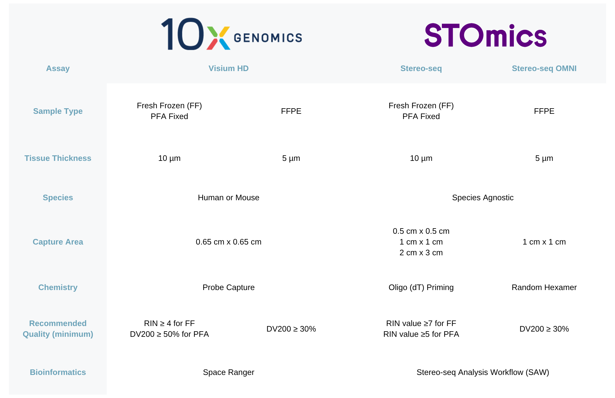
10x Genomics Visium HD is a high-definition, capture-based evolution of the original Visium spatial transcriptomics platform, capturing expression of over 18000 genes in human and muse tissues at near single-cell resolution. Its 2 μm by 2 μm barcoded tiles represent a major leap in precision compared to the 55 μm capture spots found in the standard version. Each slide offers a 6.5 mm by 6.5 mm capture area, packed with roughly 10 million barcoded features, allowing for detailed spatial profiling across human and mouse tissues.
STOmics Stereo-seq is a capture-based next-generation nanoscale spatial multi-omics platform known for its exceptional resolution and broad imaging capacity, is species agnostic, compatible with human, mouse, plant and more. With resolution down to 500 nm, it supports subcellular localization of transcripts, making it highly valuable for cellular and molecular investigations. The platform offers a range of chip sizes—from compact 0.5 cm by 0.5 cm formats to expansive 13 cm by 13 cm chips—allowing researchers to conduct high-throughput profiling of tissues as large as whole organs or embryos.
3. Choose The Right Tissue Type and Preservation Method
Not all spatial transcriptomics platforms suit every sample type, so it's crucial to match your sample with the right technology. Consider whether your tissue is fresh frozen or FFPE, and RNA quality. Admera Health supports both 10x Genomics Visium HD and STOmics Stereo-seq, which are versatile in sample compatibility and offer high-resolution gene mapping across various species. Each pushing the boundaries of resolution and tissue coverage in unique ways. They are both compatible with FFPE, fresh frozen, and fixed frozen tissues. Standard spatial samples are FFPE or OCT blocks stored under appropriate conditions and trimmed to fit processing equipment.
Fresh frozen tissues are rapidly snap-frozen to retain native morphology and RNA quality, often used when maximizing transcript integrity is a priority. Fixed frozen samples use chemical fixation (like formalin) before freezing to balance structural preservation and molecular accessibility. Meanwhile, FFPE samples offer excellent long-term storage and morphological stability, though they present challenges for RNA recovery. Embedding in OCT or paraffin and careful sectioning help maintain architecture during imaging. Staining techniques (e.g., H&E, DAPI) provide contrast for identifying features, and AI-assisted image analysis can enrich spatial mapping by extracting structural detail.
For optimal spatial transcriptomics data quality, we recommend a tissue section thickness of 5 µm for FFPE samples and 10 µm for fresh frozen or fixed frozen specimens. Ensuring high RNA integrity is critical across platforms. For Visium HD, fresh frozen samples should have a RIN ≥ 7 (acceptable RIN > 4), while fixed frozen and FFPE samples must meet a DV200 > 50%. For Stereo-seq (Stomics), the criteria are slightly different: fresh frozen tissues require a RIN > 7, fixed frozen samples a RIN > 5, and FFPE specimens should also achieve a DV200 > 50%. Adhering to these standards helps preserve transcript fidelity and spatial resolution across diverse sample types.
4. Challenges in Tissue Preparation
Proper sample collection and preparation are critical for ensuring accurate and reproducible spatial transcriptomics data. Tissue characteristics play a significant role in downstream success. Highly heterogeneous tissues such as tumors and brain may pose challenges in capturing representative regions from a single section. Conversely, tissues with variable cell density can impact signal quality—sparse samples like lymph nodes often yield low transcript counts, while densely packed tissues like pancreas may introduce signal overlap.
Preserving RNA integrity is essential, as spatial methods are particularly sensitive to degradation. Delays in freezing or fixation can substantially reduce transcript capture. Additionally, cryosectioning may introduce artifacts such as folds, tears, or ice crystal damage, compromising cellular morphology and spatial accuracy.
FFPE samples are commonly accessible but present technical hurdles due to RNA crosslinking and degradation. Moreover, fresh frozen tissues must be processed promptly—ideally within minutes to hours post-resection—to maintain viability, posing logistical challenges in clinical settings.
5. Plan for Data Analysis and Integration
Spatial transcriptomics generate large and complex datasets that require specialized analytical tools and expertise to extract meaningful insights. Key analytical steps include image registration, tissue and cell segmentation, spatial clustering, differential gene expression analysis, and data integration with complementary omics approaches such as single-cell RNA-seq or proteomics. Platforms like 10x Genomics Visium HD and STOmics Stereo-seq provide powerful software pipelines—Space Ranger and Stereo-seq Analysis Workflow (SAW), respectively—for standard processing tasks such as quality control, mapping, segmentation, clustering (e.g., Spatial UMAP), and gene expression visualization.
Due to the technical depth of these workflows, it's essential to ensure access to experienced bioinformaticians or to collaborate with service providers offering end-to-end support. Admera Health provides both standard and customized analysis options for Visium HD and Stereo-seq. Standard packages include core spatial analytics and visualization (e.g., UMAP plots, cell segmentation, tissue clustering, violin plots), while optional services enable single-cell integration and tailored analysis based on research goals.
6. Work With Experienced Partners
Spatial transcriptomics platforms involve complex experimental workflows that demand deep expertise in molecular biology, microscopy, and bioinformatics. Partnering with an experienced service provider is often the most practical and effective approach. Our lab is certified for both 10x Genomics Visium HD and STOmics Stereo-seq workflows, ensuring reliable project execution from validated requisite training and data validation.
Admera Health offers deep expertise in spatial transcriptomics across a wide range of tissue types and preservation conditions, making us a reliable partner for your research. Our team has worked with diverse species including human, mouse, cattle, rabbit, monkey, and Drosophila. We’ve successfully processed and profiled tissues from numerous organs and disease states—such as heart, kidney, spinal cord, brain, pituitary gland, liver, lung, spleen, skin, tonsil, colon/cecum, pancreatic cancer, and colon cancer—under either fresh frozen or FFPE formats. Additionally, our team of expert bioinformaticians make complex data sets generated from spatial profiling more digestible, so you can spend precious time using the analysis to make discoveries relating to your research question. This versatility enables us to support projects with complex sample requirements and tailored analytical needs.
Final Thoughts
Spatial transcriptomics is a transformative tool in functional genomics, enabling researchers to decode gene expression in context. Whether you're investigating brain development, tumor heterogeneity, or immune infiltration, thoughtful planning is essential to get the most out of your experiment.
By clearly defining your goals and aligning them with the right technologies and support, you’ll set a solid foundation for meaningful spatial insights—and avoid common pitfalls along the way.
Ready to align your next spatial transcriptomics project? Connect with one of our experts. For more information on sample submission and shipping guidelines, visit our spatial transcriptomics sample submission guideline.